
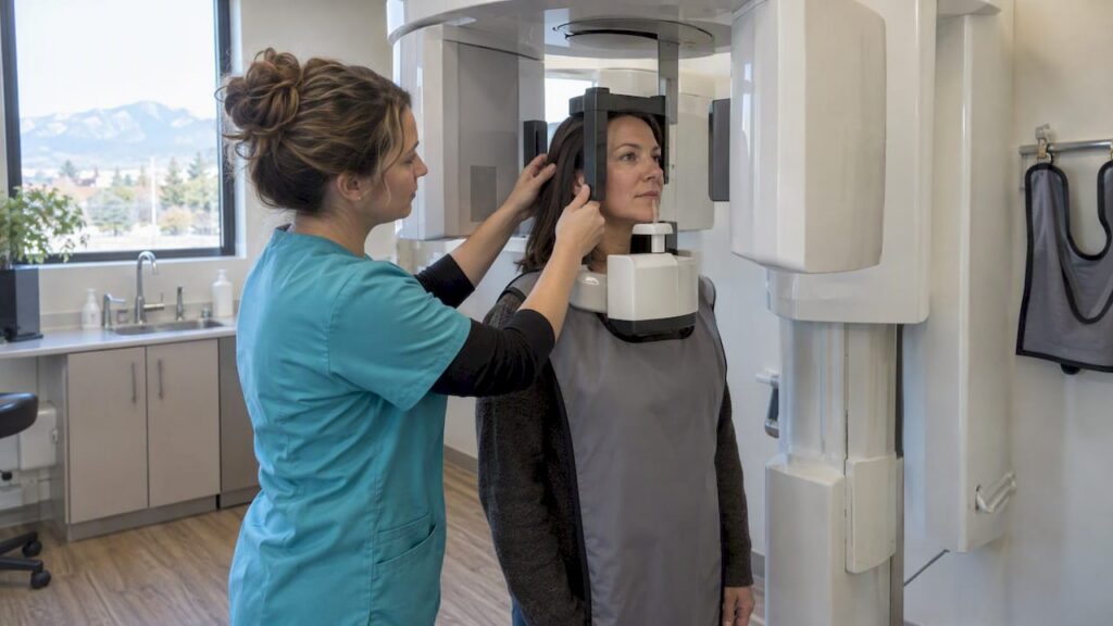
A dental CBCT scan is a three-dimensional X-ray that captures bone, teeth, and nerve pathways in a single rotation around your head. Dentists reserve it for cases where flat, two-dimensional images leave a real question unanswered, such as a hidden canal, an unclear implant site, or a suspected fracture. Professional guidance from groups like the AAE and AAOMR treats it as a targeted tool, not a routine checkup scan.
TL;DR:
- CBCT radiation doses vary significantly by FOV size, with small scans averaging 84 microsieverts and large scans reaching around 212 microsieverts, which is higher than intraoral X-rays but lower than medical CTs.
- The scan is justified only when it will meaningfully impact diagnosis or treatment, often in complex endodontic, implant, trauma, or pathology cases.
- Proper equipment maintenance, operator training on selecting minimal FOV and voxel size, and adherence to ALARA principles are essential to limit radiation exposure.
- CBCT image interpretation must consider artifacts from metal restorations and is limited in soft-tissue contrast, requiring experienced clinicians for accurate diagnosis.
- Reusing existing images and clear clinical questions help avoid unnecessary scanning and optimize patient safety and cost.
Table of Contents
- What Is a Dental CBCT and How Does It Differ From 2D X-Rays?
- How CBCT Works: Field of View, Voxel Size, and Settings
- Common Uses and Clinical Indications for Dental CBCT
- Radiation Dose and Safety: What the Numbers Show
- What to Expect During a CBCT Appointment
- Limitations, Artifacts, and Why Interpretation Matters
- How Clinicians Decide: Selection Criteria and Questions to Ask
- Regulatory and Legal Considerations: Consent and Data Privacy
- Maintenance and Calibration of CBCT Equipment
- Cost Considerations and Insurance Coverage for CBCT
- How a Modern Family Dental Practice Uses CBCT Responsibly
- Get CBCT-Informed Diagnosis and Treatment at South Littleton Family Dental
- FAQ
- Sources
What Is a Dental CBCT and How Does It Differ From 2D X-Rays?
Cone beam computed tomography sends a cone-shaped X-ray beam around your jaw in one pass, then reconstructs the data into a volumetric image made of tiny cubes called voxels. That volume can be sliced and viewed from any angle, which a flat image simply cannot do.
- Versus intraoral or panoramic X-rays: CBCT adds true depth and removes the superimposition that hides overlapping roots or canals on a 2D film.
- Versus medical CT: CBCT delivers sharper detail of bone and tooth structure at a lower dose, but its soft-tissue contrast is weaker, so it is not built for imaging organs or soft tissue masses.
This trade-off is why CBCT works well for teeth and jawbone questions and poorly for anything requiring detailed muscle or gland imaging.
How CBCT Works: Field of View, Voxel Size, and Settings
Three settings shape both image quality and radiation exposure. Field of view (FOV) ranges from small (a few teeth) to large (the full jaw and sinuses), and clinicians pick the smallest FOV that answers the clinical question, a principle reinforced in reviews of CBCT acquisition parameters. Voxel size determines sharpness.
- Small FOV: best for single-tooth endodontic or implant questions, with the lowest dose.
- Voxel size under 200 micrometers: recommended for detecting small periapical lesions or vertical root fractures, according to the same endodontic CBCT review.
- Rotation and current: switching from a 360-degree to a 180-degree rotation, or using pulsed exposure, can lower dose without sacrificing diagnostic value.
Smaller voxels sharpen fine detail, but every reduction in FOV or rotation angle also reduces the radiation a patient absorbs, a dose-saving approach documented in the same technical review.
Common Uses and Clinical Indications for Dental CBCT
CBCT earns its place in a handful of well-defined scenarios rather than as a general screening tool.
- Endodontic diagnosis: finding extra canals, investigating persistent pain after treatment, or confirming a suspected vertical root fracture.
- Implant planning: measuring bone height and width in cross-section and checking proximity to the sinus or nerve canal before surgery.
- Trauma and impacted teeth: locating fracture lines or mapping the exact position of an unerupted tooth.
- Pathology, orthodontics, and airway assessment: localizing a lesion, planning complex tooth movement, or evaluating sinus and airway anatomy.
A systematic review of CBCT’s impact on endodontic decision-making found it frequently changes diagnosis and treatment planning in trauma and complex canal cases, which is exactly why it is used selectively rather than routinely for every patient.
Radiation Dose and Safety: What the Numbers Show
Dose depends almost entirely on FOV size. A meta-analysis of published CBCT dose data reported mean adult effective doses of approximately 80 to 90 microsieverts for small FOV, around 175 microsieverts for medium FOV, and about 210 microsieverts for large FOV scans as reported in research studies, according to pooled dose research.
| FOV size | Mean adult effective dose |
|---|---|
| Small | 84 microsieverts |
| Medium | 175 microsieverts |
| Large | 212 microsieverts |
For comparison, a single intraoral X-ray delivers a small fraction of even the lowest CBCT dose, while a medical CT scan of the head typically runs far higher. Updated JADA guidance reinforces the ALARA and ALADA principles: justify every scan clinically, choose the smallest adequate FOV, and optimize machine settings before pressing the exposure button. If you are scheduled for a CBCT, it is reasonable to ask whether a child-size or low-dose protocol applies to your case and whether your dentist has already reviewed any prior imaging you have on file.
What to Expect During a CBCT Appointment
The scan itself is quick and does not require lying down; for more details on how digital imaging in dentistry enhances patient care, see Digital Imaging in Dentistry: Modern Care. You sit or stand while the machine’s arm rotates around your head, typically taking several seconds to capture the full volume.
- Before the scan: remove earrings, glasses, and any removable metal; bring dentures or retainers if your dentist wants them included in the image.
- No fasting or special prep is required beforehand.
- During the scan: you may hear a low whirring sound as the arm rotates, but there is no discomfort.
- After the scan: the raw DICOM dataset is reviewed slice by slice across axial, coronal, and sagittal planes, then summarized in a written report.
Pro Tip: Bring your current retainer, denture, or night guard to the appointment if your dentist asked for it, since removable appliances can be scanned in place to check fit against the bone.
Limitations, Artifacts, and Why Interpretation Matters
CBCT is not flawless. Metal objects such as fillings, posts, or gutta-percha from a prior root canal scatter the X-ray beam and create streaking, known as beam hardening artifact, which can obscure nearby structures.
- Metal artifacts can mask small cracks or lesions directly adjacent to a crown or post.
- Soft-tissue contrast is limited, so CBCT is a poor choice for evaluating gum tissue, tumors, or glandular structures.
- Systematic review matters: a clinician should scroll through the entire volume, not just the tooth in question, since executive guidance on CBCT interpretation stresses that incidental findings elsewhere in the scan carry diagnostic responsibility too.
When a scan involves extensive prior restorative work, an oral and maxillofacial radiologist may be asked to give a second read, since distinguishing artifact from pathology around old crowns and bridges takes practiced judgment.
How Clinicians Decide: Selection Criteria and Questions to Ask
A responsible workflow starts with a clinical exam and standard 2D imaging, and moves to CBCT only when the added detail would change the treatment plan.
- Confirm the clinical question is specific: a vague “let’s take a look” is not sufficient justification.
- Check whether a smaller FOV or lower-dose protocol fits the case.
- Verify prior images were reviewed first, since a new scan is unnecessary if an older one already answers the question.
Pro Tip: Ask your dentist what specific question the CBCT is meant to answer. A clear answer is a good sign; a shrug or a routine “we scan everyone” is a reason to ask more questions, since neither the AAE/AAOMR position statement nor ADA guidance supports scanning without documented justification.
Regulatory and Legal Considerations: Consent and Data Privacy
Before a CBCT scan, your dentist should explain why the scan is being ordered, what it will show, and what the radiation exposure involves, then document that justification in your chart. This matches long-standing ADA guidance on imaging justification, which calls for recording the clinical reasoning behind every new radiograph rather than ordering one by default.
Consent for CBCT generally follows the same standard as any other diagnostic procedure: you should understand the purpose, the general dose range relative to other imaging, and any reasonable alternatives, and you should have a chance to ask questions before the scan proceeds. If you have had a CBCT, panoramic X-ray, or medical CT scan recently, mention it. Reusing an existing image avoids duplicate exposure and reflects the same prior-image-review principle found in current radiography guidance.
Your CBCT dataset is a medical record and is handled with the same privacy protections as the rest of your dental chart, including rules around who can access, transfer, or request copies of your imaging. Scans are typically stored in DICOM format, which can be shared between your general dentist, an endodontist, or an oral surgeon when a referral is needed, though such transfers should happen through secure channels and with your authorization. If a scan is sent to a specialist for a second opinion or a surgical referral, ask how the image will be transmitted and how long it will be retained. These are reasonable questions at any dental office, and a practice that handles imaging routinely should have a clear answer.
Maintenance and Calibration of CBCT Equipment
Image quality and radiation safety both depend on equipment that is properly maintained, not just correctly operated. CBCT units require periodic calibration to confirm that the X-ray output matches the settings selected on the control panel, since a drift in calibration can mean a patient receives a higher dose than the protocol intended while producing a lower-quality image.
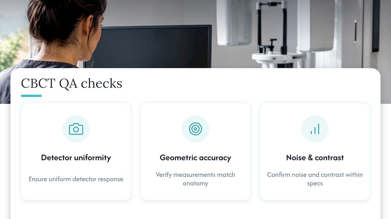
Routine quality assurance typically includes checking detector uniformity, verifying geometric accuracy so measurements on the scan reflect real anatomical dimensions, and confirming that image noise and contrast remain within the manufacturer’s specifications. Offices that use CBCT regularly track these checks on a schedule, often tied to manufacturer recommendations and applicable state or federal radiation safety rules for medical imaging equipment.
Beyond scheduled calibration, staff operating the machine need training on selecting the correct FOV and voxel size for each clinical situation, since equipment that is perfectly calibrated still produces an unnecessarily high dose if the operator defaults to the largest FOV out of habit. Reviewing dose output periodically, sometimes against institutional reference levels, helps a practice confirm it is staying consistent with the ALARA approach described in current radiation safety guidance rather than letting default settings creep upward over time.
Cost Considerations and Insurance Coverage for CBCT
CBCT pricing varies by office, region, and the complexity of the scan, since a small single-tooth FOV involves less imaging time and data than a full-arch large FOV capture. Because pricing is set at the practice level and tied to the specific clinical indication, there is no single universal number that applies to every patient or every scan type.
Dental insurance coverage for CBCT is inconsistent. Some plans cover a portion of the cost when the scan is tied to a covered procedure, such as implant placement or root canal therapy, while others classify CBCT as a separate diagnostic charge that is only partially reimbursed or not covered at all. Medical insurance occasionally covers CBCT when it is used for airway or sinus evaluation tied to a medical diagnosis, though this varies by carrier and policy.
Before scheduling, it is worth asking your dental office whether CBCT is billed separately from the associated treatment and whether your specific insurance plan has a history of covering it for your indication. Offices that handle CBCT regularly can usually tell you in advance whether a claim is likely to be approved, which avoids a surprise charge after the scan is already done.
How a Modern Family Dental Practice Uses CBCT Responsibly
We fold CBCT into personalized care plans rather than treating it as a default step, using it when three-dimensional detail will genuinely change a diagnosis or a surgical plan. For same-day emergency exams, a targeted field of view is selected to match the clinical question, whether that means mapping an implant site or confirming a suspected fracture, and findings are discussed with the patient before deciding on next steps.
— Admin
Get CBCT-Informed Diagnosis and Treatment at South Littleton Family Dental

When a tooth problem is hard to pin down from a standard exam, CBCT-informed imaging gives our team the detail needed to plan treatment with confidence instead of guesswork. We use it to support root canal therapy when a canal system is unclear, to assess bone before dental implants, and to evaluate urgent cases during same-day emergency exams. Our advanced imaging technology lets us scan only when it will change your care plan, keeping exposure and cost in line with what each case actually needs.
If you are dealing with persistent tooth pain, considering implants, or facing a dental emergency, visit our general dentistry page to see appointment options, or call our Littleton, CO office directly to ask about same-day availability. Bring any prior X-rays or scans you have, since reviewing existing images first is part of how we decide whether a new CBCT is warranted.
This article is general information, not a substitute for advice from a qualified doctor. Consult a qualified healthcare professional about your own circumstances before acting on anything here.
FAQ
Does a CT scan show dental problems?
Yes, both medical CT and dental CBCT can reveal dental problems such as fractures, impacted teeth, and bone loss around the jaw. CBCT is generally preferred for tooth-specific diagnosis because it offers sharper detail of bone and tooth structure at a lower radiation dose than a full medical CT scan.
Do you need CBCT for a root canal?
Not for every root canal. CBCT is typically reserved for cases with persistent pain after treatment, a suspected extra canal, or a possible vertical root fracture, since a systematic review of endodontic cases found it most often changes treatment planning in these more complex situations rather than in routine procedures.
What are the risks of CBCT?
The main risk is exposure to ionizing radiation, though doses are generally low, averaging around 84 to 212 micrograys depending on field of view size according to pooled dose data. Clinicians reduce this risk further by choosing the smallest field of view and lowest settings that still answer the clinical question, following current safety guidance.
How does CBCT work?
A CBCT machine rotates a cone-shaped X-ray beam around your head in a single pass, capturing hundreds of images that are reconstructed into a three-dimensional volume made of tiny voxels. This volume can then be viewed from any angle or sliced into cross-sections, which lets a dentist examine bone and tooth structure in a way a flat X-ray cannot.
Sources
- Cone-beam computed tomography in endodontics: from the specific technical considerations of acquisition parameters and interpretation to advanced clinical applications (PMC)
- Effective dose of dental CBCT—a meta analysis of published data (PubMed)
- Optimizing radiation safety in dentistry – The Journal of the American Dental Association (JADA)
- Cone Beam Computed Tomography Impact on Diagnosis, Treatment Planning and Confidence in Endodontic Decision-Making: A Systematic Review (PubMed)