
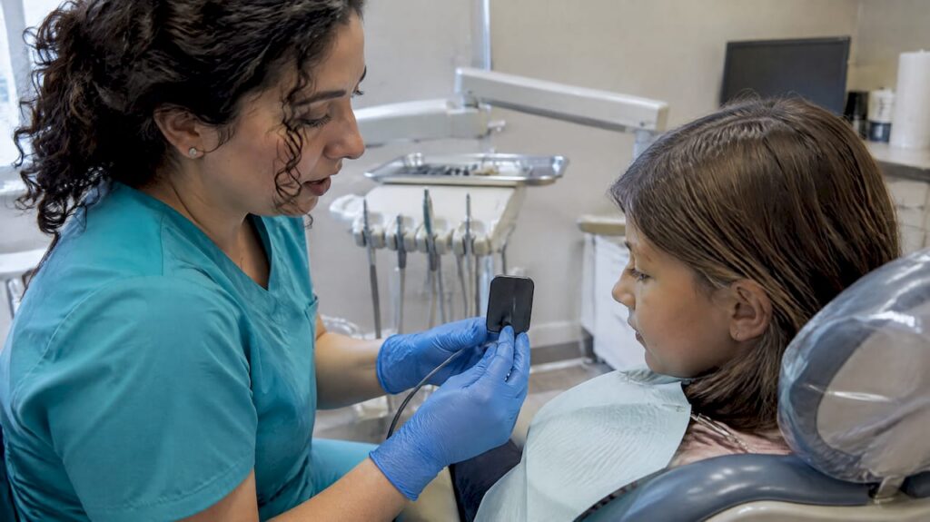
Modern dental X-rays are low-dose and considered safe when a dentist orders them for a clear clinical reason. Professional guidance built on the ALARA principle, “as low as reasonably achievable,” shapes how often images are taken and at what dose. Pregnant patients and children receive specific precautions on top of these standard safeguards.
TL;DR:
- Adults at low risk may wait two to three years between bitewing images; higher risk patients may need them every six to eighteen months.
- CBCT scans can expose patients to many times more radiation than standard intraoral images, so dentists reserve them for cases where 3D detail changes treatment.
- Ask whether the office uses digital sensors and rectangular collimation, which reduce dose by using sensitive detectors and limiting the beam to the target area.
- Tell your dentist about pregnancy, a child’s age, prior scans, or cancer radiation therapy so the team can weigh necessity, timing, and lower dose options.
Table of Contents
- How much radiation do common dental X-rays give?
- Types of dental X-rays and when each is used
- How often are dental X-rays needed?
- Safety measures dentists use to minimize radiation
- Pregnancy and children’s safety considerations
- Common patient questions and side effects
- How South Littleton Family Dental applies safety best practices
- Who should avoid or limit dental X-rays beyond pregnancy and children
- Technological advancements improving dental X-ray safety
- Patient preparation and communication to reduce anxiety
- Practical takeaway for patients and parents
- South Littleton Family Dental: safe imaging and patient care
- FAQ
- Sources
How much radiation do common dental X-rays give?
Dose depends heavily on which type of image your dentist takes. Bitewing and periapical X-rays, the small images used to check for cavities or look at a single tooth root, sit at the low end of the scale. Panoramic X-rays, which capture the entire jaw in one sweep, use a somewhat higher dose but remain modest. Cone-beam computed tomography (CBCT), used for 3D views of bone and implant planning, can deliver a substantially higher dose than either of those options, since it captures far more anatomical detail.
- Bitewing and periapical images: very low dose, often comparable to brief environmental exposure.
- Panoramic images: higher than bitewings but still considered low-dose overall.
- CBCT scans: can be many times higher than a standard intraoral image, reserved for specific diagnostic needs.
A single dental X-ray often delivers less radiation than the background radiation you absorb in a single day. That comparison helps put routine dental imaging into perspective against the radiation we encounter simply by existing on Earth.
Dental radiographs also make up a small share of a person’s total imaging exposure over a lifetime compared with medical scans like chest CTs or abdominal imaging, which involve much larger areas of the body and higher doses per exam; for more on chest imaging requirements and safety, see Do I Need a Chest X-Ray for My Immigration Medical Exam?
Types of dental X-rays and when each is used
Your dentist picks the type of X-ray based on the specific question that needs answering, not as a routine formality.
- Bitewing X-rays show the crowns of upper and lower teeth together, ideal for spotting cavities between teeth.
- Periapical X-rays capture a whole tooth from crown to root tip, used to check for infections, abscesses, or bone loss around a single tooth.
- Panoramic X-rays give a wide view of the jaws, teeth, sinuses, and joints, useful for wisdom tooth evaluation, orthodontic planning, or spotting cysts and tumors.
- CBCT scans produce a 3D image of bone and tooth structure, used mainly for implant planning, complex root canal cases, or surgical assessment.
Because CBCT carries a higher dose, it’s reserved for situations where 3D detail genuinely changes the treatment plan rather than used as a default option.
How often are dental X-rays needed?
There’s no universal schedule that applies to everyone. The FDA’s guidance on patient selection calls for imaging only after a clinical exam and review of dental and medical history, tailored to each patient’s age and risk factors rather than a fixed calendar.
- New patients typically receive a baseline set of X-rays to establish a starting point for future comparison.
- Low-risk adults with good oral health and no history of cavities may go two to three years between bitewing X-rays.
- Higher-risk patients, including those with a history of frequent cavities, gum disease, or certain health conditions, may need images every six to eighteen months.
- Children often need more frequent monitoring while teeth and jaws are developing, though intervals still depend on individual risk.
Updated ADA recommendations released in 2026 reinforce that radiographs should be ordered only when clinically necessary, following ALARA at every step. A dentist who reviews your prior images before ordering new ones, rather than defaulting to a routine retake, helps avoid exposure that adds nothing to your care.
Safety measures dentists use to minimize radiation
The ALARA principle drives a set of concrete practices in the treatment room, not just a policy statement. Dentists reduce dose through equipment choices and technique rather than relying on a single safeguard.
- Digital sensors or F-speed film instead of older, less sensitive film, which can substantially cut patient dose.
- Rectangular collimation, which narrows the X-ray beam to the area actually being imaged instead of a wider circular field.
- Proper positioning with receptor holders to avoid retakes caused by blurry or misaligned images.
- Adequate source-to-skin distance and appropriate kVp settings calibrated to the equipment.
Shielding practices have shifted in recent years. Current professional guidance notes that thyroid collars and lead aprons aren’t routinely necessary for intraoral, panoramic, or CBCT imaging, since collimation and modern equipment already limit stray radiation effectively. Some practices still offer a thyroid collar on request or for specific cases, which is a reasonable option rather than a requirement.
Pro Tip: Ask your dental office whether they use digital sensors and rectangular collimation. Those two details tell you a lot about how seriously a practice takes dose reduction.
Pregnancy and children’s safety considerations
Dental X-rays target the mouth and jaw, not the abdomen, so fetal exposure is typically negligible. Research on dental radiography during pregnancy confirms that imaging of the teeth doesn’t expose an unborn baby to meaningful radiation, though telling your dentist if you’re pregnant or think you might be lets the team confirm the exam is necessary and apply standard protocols.
Children receive their own set of adjustments, since their developing tissues are more sensitive to radiation.
- Equipment settings are scaled down for smaller mouths and shorter exposure times.
- Images are limited to views that will actually change treatment decisions.
- Nonessential imaging can reasonably be delayed when a child’s oral health doesn’t require it right away.
CDC guidance for parents stresses that the benefits of needed imaging usually outweigh the risks, while recommending dose-adjusted settings and avoiding unnecessary repeat exams in children. Asking whether a practice adjusts exposure for your child’s size is a reasonable question for any parent.
Common patient questions and side effects
Patients often hear terms like “leakage radiation” without a clear explanation. Leakage radiation refers to small amounts of radiation that escape the X-ray tube housing outside the main beam; modern equipment is built with shielding that keeps this to a minimal level, well within safety standards.
- Routine dental radiographs produce no physical side effects such as pain, burns, or lingering sensations.
- Long-term risk from occasional, clinically necessary dental X-rays is considered very low given the small doses involved.
- Four bitewing images together still represent a low overall dose, roughly in the range of routine background exposure over a short period.
- A properly functioning, well-maintained machine is a key factor in keeping both scatter and leakage radiation low.
For deeper technical detail on dose comparisons and equipment standards, the FDA’s dental radiography resources offer additional background.
How South Littleton Family Dental applies safety best practices
We built our imaging approach around the same selection criteria and dose-reduction habits described above. Our advanced technology includes digital sensors, and we review a patient’s prior images before deciding whether a new X-ray adds anything to the care plan. We pair that with same-day emergency availability and individualized care plans, so imaging decisions always connect back to a specific diagnostic question rather than a routine checkbox.
Who should avoid or limit dental X-rays beyond pregnancy and children
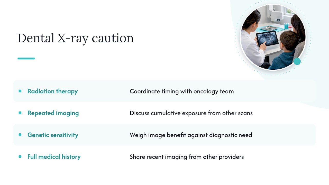
Pregnancy and childhood get most of the attention, but a few other situations call for a closer conversation with your dentist before imaging. Patients undergoing radiation therapy for head or neck cancer may already have accumulated significant radiation exposure in that area, so your dentist and oncology team may coordinate on timing and necessity of additional dental images. People with a documented history of frequent diagnostic imaging across multiple specialties, such as repeated CT scans for an unrelated condition, may also benefit from a conversation about cumulative exposure, even though dental doses are individually small.
Patients with certain genetic conditions that increase radiation sensitivity, though uncommon, should mention this history so the dental team can weigh the benefit of an image against the diagnostic need more carefully. There’s no blanket condition that rules out dental X-rays entirely for most adults; the more common scenario is simply adjusting frequency or substituting a lower-dose image type when a clinical history suggests extra caution is reasonable.
The practical takeaway is the same across all these cases: share your full medical history, including recent imaging from other providers, so your dentist can factor it into the decision. A dentist who asks about your broader imaging history before ordering a new X-ray is applying the same selection principle that governs pregnancy and pediatric care, just extended to anyone with a reason for added caution.
Technological advancements improving dental X-ray safety
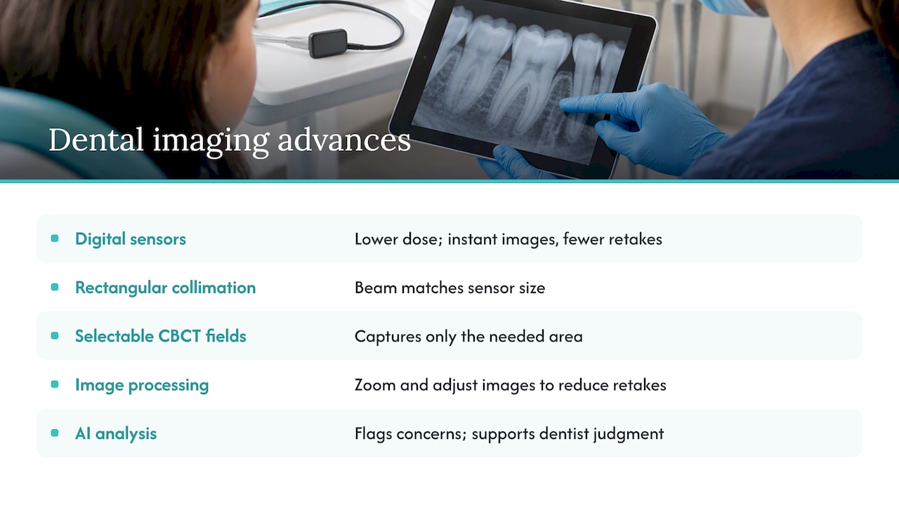
Dental imaging equipment has changed considerably from the film-based systems many adults remember from childhood visits. Digital sensors replaced traditional film in most practices, cutting radiation dose substantially while also producing images instantly, which means fewer retakes from blurry or poorly positioned film.
Rectangular collimation, now standard in updated equipment, narrows the X-ray beam to match the size of the digital sensor rather than exposing a wider circular area of tissue that isn’t being imaged. Panoramic and CBCT machines have also improved: newer CBCT units offer selectable fields of view, so a dentist can capture just the specific area needed, such as a single implant site, rather than scanning the entire jaw at a higher dose than necessary.
Image processing software now allows dentists to zoom, adjust contrast, and enhance digital images after the fact, which reduces the need for a repeat exposure when the first image doesn’t show enough detail. Some practices also use AI-assisted analysis tools to help flag areas of concern on an image, supporting the dentist’s judgment rather than replacing it. Together, these advancements mean today’s equipment can produce clearer diagnostic images at a lower dose than equipment from even a decade ago.
Patient preparation and communication to reduce anxiety
A short conversation before the X-ray often does more to ease anxiety than anything else. Dentists who explain why a specific image is needed, what it will show, and how long the process takes give patients a clear sense of control over an otherwise unfamiliar procedure.
Simple preparation steps help too: removing jewelry or metal objects near the head and neck, wearing the provided protective apron if offered, and sitting still for the few seconds each image requires. For patients with dental anxiety, letting the team know in advance allows them to explain each step before it happens, rather than starting the machine without warning.
Children in particular benefit from a brief, age-appropriate explanation, sometimes paired with letting them see the sensor or hold a model, before the actual image is taken. Patients who ask questions, such as how many images will be taken or whether prior X-rays are already on file, tend to feel more at ease throughout the visit. Good communication doesn’t change the physics of the exposure, but it does change how the experience feels, which matters for patients who’ve avoided dental visits specifically because of X-ray anxiety.
Practical takeaway for patients and parents
Dental X-rays are safe when they answer a real clinical question, not when they’re routine for routine’s sake. Three questions are worth asking at any visit: Is this image necessary right now? Do you have my prior X-rays on file? What dose-reduction steps will you use? A dentist willing to answer all three is practicing good imaging medicine.
— Admin
South Littleton Family Dental: safe imaging and patient care
We bring the same dose-reduction practices covered throughout this article into every exam room: digital sensors, rectangular collimation, and a review of your prior images before ordering anything new. Our general dentistry visits include exams and X-rays guided by these selection principles, and our approach to X-ray frequency follows the same individualized criteria described above rather than a fixed schedule.

- Emergency exams are available when a dental issue can’t wait.
- Questions about a specific X-ray or a child’s imaging needs are welcome before booking.
If you have questions about an upcoming exam or want to schedule a visit, reach out through our general dentistry page to get started.
This article is general information, not a substitute for advice from a qualified doctor. Consult a qualified healthcare professional about your own circumstances before acting on anything here.
FAQ
How much radiation is in four bitewing X-rays?
Four bitewing X-rays together still represent a low overall dose, often comparable to the natural background radiation a person receives in a single day. The exact figure varies by equipment and technique, which is one reason digital sensors and proper collimation matter.
What is leakage radiation?
Leakage radiation is a small amount of radiation that escapes the X-ray tube housing outside the primary beam aimed at your mouth. Modern dental equipment is shielded to keep this leakage within recognized safety limits, so it isn’t a meaningful source of extra exposure during a routine exam.
What are the side effects of a dental X-ray?
Routine dental X-rays don’t cause physical side effects like pain, burns, or lingering symptoms. The main consideration is cumulative radiation exposure over a lifetime, which is why dentists limit imaging to what’s clinically necessary and follow the ALARA principle.
Are digital dental X-rays safer than traditional film?
Yes, digital sensors generally require less radiation than older film-based systems to produce a usable image. Switching to digital receptors or faster film speeds has been shown to meaningfully reduce patient dose compared with older D-speed film.
How often should I get dental X-rays?
Frequency depends on your individual risk factors, age, and dental history rather than a fixed rule for everyone. Low-risk adults may go two to three years between bitewing X-rays, while higher-risk patients or children may need imaging more often, based on a dentist’s clinical evaluation.
Sources
- MouthHealthy — X-rays
- FDA — Selection of patients for dental radiographic examinations
- ADA press release — New recommendations confirm dental X-rays most effectively used in moderation
- CDC — Facts about radiation in healthcare for parents