
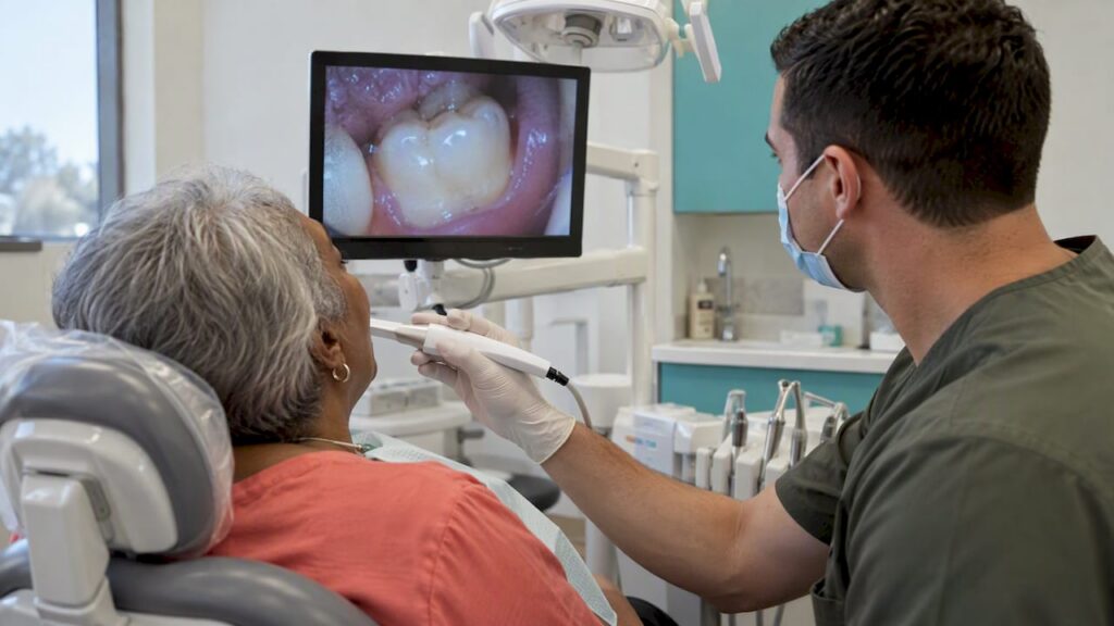
An intraoral camera is a small, pen-shaped camera that your dentist passes around your mouth to show a magnified live picture of your teeth and gums on a screen. The main benefits are simple: you can see your own mouth, problems tend to be spotted earlier, treatment recommendations are easier to understand, and your images are saved so changes can be tracked from one visit to the next. It is a helper for the dentist’s judgment, not a replacement for it, and it does not replace X-rays. Below we explain how it works, what the research says, where it falls short and what to ask your dentist.
TL;DR
- An intraoral camera shows a magnified, real-time view of your teeth and gums on a monitor, so you can see what the dentist sees.
- Research links it to better patient understanding, stronger motivation and better acceptance of recommended treatment, plus better records over time.
- It is useful for cracks, plaque, tartar, worn spots and soft-tissue changes, but it only shows surfaces. It does not see between teeth or inside bone the way X-rays do.
- It uses no radiation, and you can ask how your images are stored and who can see them.
- Ask your dentist to show you your images during the exam, point out the problem area and explain the options.
Table of Contents
- What an intraoral camera is
- The benefits of an intraoral camera, one by one
- What an intraoral camera can’t do
- Camera, scanner or X-ray: which is which?
- Your images and your privacy
- Who benefits most
- Questions to ask your dentist about the camera
- Our perspective
- See what we see at your first visit
- FAQ
- Sources
What an intraoral camera is
The name is more technical than the tool. “Intra” means inside, “oral” means mouth. The camera is a slim handheld wand with a tiny lens and light at the tip. The dentist or hygienist holds it close to a tooth, and the image appears on a chairside monitor.
The U.S. Food and Drug Administration describes this kind of device plainly. In its 2009 clearance summary for one widely sold model, it is a chairside system that captures and stores still and video images and shows magnified digital color views of the inside or outside of the mouth on a monitor or PC. The same document lists it as a Class I device, the FDA’s lowest-risk category, meant to help assess a patient’s overall oral health.
Inside the wand there is an image sensor, usually a CCD or CMOS chip, the same family of sensors found in digital cameras. A 2019 review explains that the sensor converts light into electronic signals that software turns into an image on the screen. That is all there is to it: light, a sensor, software and a monitor.
What it looks like during your visit
Most patients barely notice it. The wand is about the size of a thick pen and the tip is small. The dentist moves it slowly along the surfaces of your teeth, the gumline and sometimes the tongue, cheeks and palate. You do not need to bite on anything or hold still for long, and there is no flash. Pictures can be frozen on the screen, and some are saved to your chart.
Good images depend on technique. A 2021 report on intraoral photography notes that adequate lighting and attention to camera angle are what make pictures readable. This is one reason a trained clinician holds the camera, and why the picture is only as useful as the person interpreting it.
The benefits of an intraoral camera, one by one
The pages that rank for this topic tend to list the same handful of advantages. Here is what each one means in practice, and what the evidence actually supports.
1. You can see the problem yourself
This is the biggest benefit for most patients. A dentist can say “you have a crack in that filling” and you have to take their word for it. With a camera, the crack is on the screen, enlarged many times.
A 2026 study in the British Dental Journal concluded that camera images can help patients see conditions such as plaque, understand their oral health and take part in shared decisions about treatment. A 2020 American Dental Association blog post written by a dentist describes the same effect: showing patients real-time images of heavy buildup or a cracked filling helps them understand what the dentist sees, which can improve treatment acceptance.
That matters most for problems that do not hurt. Early decay, a hairline crack or gum inflammation often cause no pain, so a recommendation can feel abstract. A picture makes it concrete.
2. Problems can be caught earlier
A mouth mirror and good overhead light go a long way, but a camera adds magnification and reaches the back molars, the tongue side of lower teeth and other tight spots. The British Dental Journal study describes modern cameras as ergonomically designed and useful for finding cracks, abrasions, erosion, periodontal disease, plaque and calculus, and for locating canal openings during root canal work.
Earlier detection usually means a smaller, simpler repair. A small chip or early worn spot may be treated with a conservative filling, while the same problem found later can need a crown. We cannot promise any particular outcome, but finding things sooner is the whole point of regular exams.
3. Patients are more likely to follow through
There is real data here. A 2019 study reported by the U.S. National Library of Medicine looked at a dental school clinic and found that using intraoral cameras significantly increased the completion rate of treatment-planned posterior crowns. In plain terms, patients who saw their images were more likely to go ahead with the crown that was recommended.
The 2019 review mentioned earlier makes a related point: clear visual education before, during and after treatment can increase patient compliance and motivation. Seeing the buildup along your gumline is a stronger reminder to floss than a lecture.
A fair reading: a camera does not decide for you, and a good dentist uses it to inform, not to pressure. The goal is that you understand the choice and make it with the facts in front of you.
4. A visual record builds over time
An image saved today can be compared with one taken next year. That makes it easier to tell whether a dark spot is stable, a worn edge is getting worse or a patch of gum is improving after a deep cleaning. The British Dental Journal study lists better documentation and the ability to track changes in oral health over multiple visits among the camera’s benefits, along with simpler remote consultations.
Records also help when something needs a second opinion, a specialist referral or an insurance review. A clear photo of a fractured tooth says more than a paragraph of notes.
5. It is comfortable and uses no radiation
The camera only looks. It does not press hard on anything, and it produces light, not X-rays. The FDA clearance summary reports that non-clinical testing of the hardware, software and assembled system showed it was safe and effective for its intended use. For children, anxious patients and anyone with a strong gag reflex, a small wand is usually easier to tolerate than some other tools. If you are nervous, ask the team to tell you before the camera goes in, and to pause whenever you need it.
6. Better conversations between you and your dentist
The American Dental Association has made the point that digital tools are helpful adjuncts to a practitioner’s knowledge, helping to find answers and communicate them to patients. The wording matters. The tool supports clinical expertise. It does not replace it.
The British Dental Journal study adds a condition that applies to every dental technology: effective communication and trust between patient and dentist are what make people accept the tool in the first place. A camera works best when the person holding it explains what you are looking at in plain language.
What an intraoral camera can’t do
A camera is not a complete exam. Being honest about its limits is part of using it well.
It only shows surfaces. The camera sees what light can reach: the visible parts of teeth and gums. It cannot show decay between tightly touching teeth, infection at the root tip, bone loss around a tooth or the position of unerupted teeth. Those need X-rays or other imaging. A 2017 study notes that two-dimensional radiographs are commonly used to evaluate hard tooth structures beneath the surface, which is exactly the territory a surface camera cannot cover.
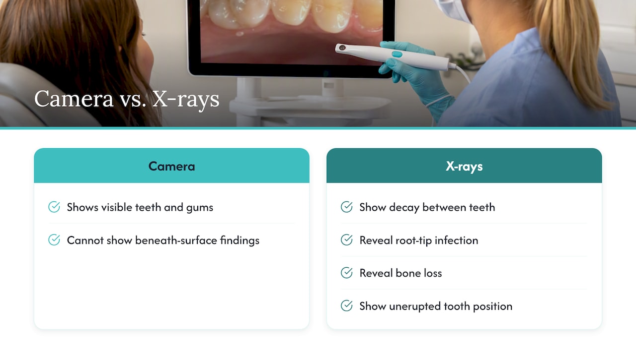
It is not a diagnosis on its own. A picture of a dark spot does not tell the dentist whether it is a stain, an old filling margin or active decay. The dentist combines the image with a hands-on exam, your history and, when needed, X-rays.
It is not the best tool for photographing lesions. The 2021 report on intraoral photography does not recommend intraoral cameras for photographing oral lesions, because they can produce a narrow, magnified, blurry image with no neighboring structures for reference. For tracking a sore, patch or lump, standardized photographs that include surrounding anatomy are essential. The BDJ study does say cameras can help screen for soft-tissue changes, including ulcers and early signs of oral cancer, so the camera has a role in the screening exam, but anything suspicious should be evaluated and documented properly by your dentist or a specialist.
Camera, scanner or X-ray: which is which?
Patients often mix these three up, and the search results show it: people who look up the camera also ask what an intraoral scan is.
| Tool | What it does | What it shows | Radiation |
|---|---|---|---|
| Intraoral camera | Takes live video and still photos | Surface of teeth, gums, soft tissue | None |
| Intraoral scanner | Builds a 3D digital model of your teeth, often for aligners, crowns or other restorations | Shape and position of teeth and bite | None |
| Dental X-ray | Passes a very low dose of radiation through teeth and bone | Cavities between teeth, root tips, bone levels | Low dose |
The camera is for looking and explaining. The scanner is for measuring and designing. The X-ray is for seeing what is hidden. A thorough exam often uses more than one, and each answers a different question.
Some researchers are testing camera-based systems that use near-infrared light to detect problems under the surface. The 2017 study found such a system could detect subsurface dental conditions, and noted that non-ionizing imaging is a major draw for patients worried about radiation. That is promising, but it is a different device from the standard visible-light camera most offices use today.
If X-ray exposure is your worry, you do not have to skip them. Ask about digital sensors and how often you really need images; our page on dental X-ray safety for families covers the questions worth asking.
Your images and your privacy
Photos of your mouth are part of your health record. The British Dental Journal study points out that patients may have privacy concerns about camera images, which makes informed consent and clear communication important for trust and data security.
It is reasonable to ask:
- Are my images saved to my chart, and for how long?
- Who can see them besides my dental team?
- Will you ask me before using them for anything other than my care, such as teaching or sharing with a specialist?
A good office answers these easily.
Who benefits most
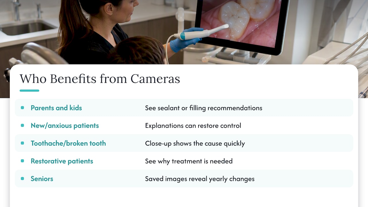
Parents and kids. Children often cannot describe what bothers them. Letting a child see a “tooth movie” on the screen takes away some mystery and can turn an exam into something curious instead of scary. Parents also see exactly where a sealant or filling is being recommended.
First-time and anxious patients. Fear often comes from not knowing what is happening. A screen that shows each step, with a calm explanation, gives you back some control.
Patients with a toothache or broken tooth. When you walk in with pain, a close-up of a cracked cusp or damaged filling shows the cause quickly and helps you weigh options on the same day. Our emergency dentistry page explains what to do when you cannot wait.
Anyone facing a crown, bridge, implant or other restorative work. These treatments cost real money and time. Seeing why a tooth needs one is the fairest basis for deciding. You can read about options on our restorative dentistry page.
Seniors. Worn fillings, gum recession and ill-fitting dentures are easier to understand when you can see them, and saved images make changes obvious from year to year.
Questions to ask your dentist about the camera
You do not need to be an expert. These questions get you the most from it:
- Can you show me the image of the tooth you are concerned about?
- What exactly am I looking at: decay, a crack, buildup or an old filling?
- Is this urgent, or can it be watched? What happens if we wait?
- Do I also need an X-ray to confirm this?
- Can you save this image so we can compare it at my next visit?
- What are my options, and what does each cost with and without insurance?
If you leave an exam unsure what the dentist found, the visit did not do its job. A camera is just a way to make the explanation easier.
Our perspective
We see dentistry as a conversation first and a procedure second. Many of the people who come to us are parents juggling a schedule, adults who are in pain and want answers, seniors weighing restorations, and people who have avoided the dentist for years because they felt rushed or talked down to. Clarity and detail are what calm those fears.
That is how we think about any imaging tool, including the intraoral camera. We want you to understand your own mouth, not nod along. When you can see a problem, you can ask better questions, decide with confidence and say no to what you do not need. We also want you to hear the limits: a picture is one part of the exam, and it does not replace the dentist’s hands-on evaluation, X-rays when they are needed or a care plan built around you.
If you want to know more about the technology we bring into the office, our advanced technology page is the place to start. And if a team is not willing to show you what they see, that is worth noticing.
— Admin
See what we see at your first visit
We offer general exams and cleanings, same-day emergency care, restorative work, cosmetic treatment and children’s dentistry in Littleton, CO. If you are new to us, start with our new patient special, or look at general dentistry to see what a routine visit includes. Tell us at the front desk if you are nervous. We will go at your pace and explain each step.
FAQ
What is an intraoral camera?
It is a small, pen-shaped digital camera that a dentist or hygienist uses inside your mouth. It sends a magnified live image to a monitor and can save still photos or video. The FDA classifies standard models as low-risk Class I devices intended to help assess oral health.
Does an intraoral camera hurt?
No. The wand is small and only needs to be held near your teeth and gums, with no pressure or flash. Most people find it more comfortable than being told about a problem they cannot see. If you have a sensitive tooth or a strong gag reflex, tell the team and they can adjust.
Does an intraoral camera replace dental X-rays?
No. A camera shows only surfaces you can see. X-rays show what is hidden, such as decay between teeth, infection at the root and bone levels. Dentists use the two together, and your dentist decides when X-rays are needed based on your history and risk.
Can an intraoral camera detect oral cancer?
It can help in a screening exam by letting the dentist look closely at soft tissue for ulcers, patches and other changes, according to a 2026 British Dental Journal study. It cannot diagnose cancer. Anything suspicious needs a proper clinical evaluation and, if necessary, a referral to a specialist for testing.
Are my camera images private?
They are part of your dental record, so they should be handled like the rest of it. Ask the office how images are stored, who can access them and whether they will ask your permission before using them for anything beyond your care.
Sources
- FDA 510(k) summary K091186, intraoral camera, 2009
- Clinical applications of intraoral camera to increase patient compliance, 2019
- Intraoral Photography Recommendations for Remote Risk Assessment, 2021
- Intra-oral camera utilisation: a multi-perspective study, British Dental Journal, 2026
- Intraoral Camera Use in a Dental School Clinic, 2019
- Dental Practice Success, American Dental Association, 2016
- Implementing new technology into an older practice, ADA New Dentist Blog, 2020
- Near-Infrared Imaging for Detecting Caries and Structural Deformities, 2017