
Lost gum tissue does not grow back on its own. Gum recession treatment focuses on stopping further loss, reducing sensitivity, and, when the recession is significant enough, surgically restoring coverage through grafts or flap procedures. The sooner you address it, the more options you have.
Your two main clinical paths:
-
Conservative (non-surgical): Correcting brushing technique, professional scaling and root planing (SRP), desensitizing agents, and behavior changes that remove the cause. This stops progression and eases symptoms but does not restore lost tissue volume.
-
Surgical: Procedures such as the coronally advanced flap (CAF), subepithelial connective tissue graft (CTG), modified coronally advanced tunnel (MCAT), free gingival graft (FGG), acellular dermal matrix (ACDM), collagen matrix (CM), and enamel matrix derivative (EMD) adjuncts. These can restore root coverage with predictable results when the case is selected carefully.
When to call your general dentist: mild sensitivity, a tooth that looks slightly longer than it used to, or a routine checkup where you want recession measured and monitored.
When to ask for a periodontist referral: multiple teeth affected, rapidly progressing recession, significant root exposure, root decay, or any case where surgical grafting is being considered.
Table of Contents
-
What causes receding gums and what symptoms should you watch for?
-
How do dentists diagnose recession and when should you seek care?
-
What non-surgical treatments can stop recession from getting worse?
-
What should you expect after gum surgery: recovery, risks, and costs?
-
When recession coexists with other problems: NCCLs, orthodontics, and bite issues
-
How to prevent further recession and protect your gum health long term
-
What the evidence actually says about treating gum recession
What is gum recession and how common is it?
Gingival recession is the clinical term for what happens when the gum margin migrates apically, meaning it moves down and away from the crown of the tooth, exposing the root surface. That exposed surface is covered in cementum, a softer tissue than enamel, which has no protective outer layer. Cementum is more vulnerable to acid erosion, bacterial plaque, and abrasion, which is why exposed roots tend to feel sensitive and are more prone to decay than the crown above the gumline.
The clinical consequences stack up quickly:
-
Tooth sensitivity: Cold liquids, air, and sweet foods trigger sharp, short pain from exposed dentinal tubules.
-
Root caries: Exposed cementum decays faster than enamel; untreated root cavities can progress to the pulp.
-
Non-carious cervical lesions (NCCLs): Notch-shaped wear defects at the gumline, often caused by abrasion or erosion, frequently coexist with recession.
-
Aesthetic change: Teeth appear longer; dark triangles may form between teeth.
-
Progression to tooth loss: When recession accompanies uncontrolled periodontal disease, attachment loss continues until the tooth becomes mobile.
Recession is far more common than most people realize. The American Academy of Periodontology notes that gum disease, one of the primary drivers of recession, affects a large share of American adults. Recession itself increases with age and is frequently found even in patients with otherwise healthy mouths, particularly on the lower front teeth and upper canines.
What causes receding gums and what symptoms should you watch for?
Symptoms you may notice first
-
A tooth that looks noticeably longer than its neighbors
-
Sharp sensitivity to cold water, cold air, or sweet foods
-
A visible yellowish or darker root surface below the gumline
-
A notch you can feel with your tongue at the base of a tooth
-
Bleeding when brushing or flossing
-
Occasional aching or tenderness along the gumline
Why gums recede: the main causes
Aggressive brushing is the single most common non-disease cause. Scrubbing with a medium or hard brush, or using a horizontal sawing motion, wears away both gum tissue and cementum over years. Many patients are surprised to learn their diligent brushing is the problem.
Periodontal disease drives recession through bacterial infection that destroys the connective tissue and bone supporting the tooth. This is the cause most likely to produce deep pockets, bone loss, and eventual mobility alongside the recession.
Thin tissue biotype and genetics play a real role. Patients with naturally thin, delicate gum tissue and thin underlying bone are predisposed to recession even with perfect hygiene. There is not much you can do about your biotype, but knowing it helps your dentist plan preventively.
Bruxism and occlusal trauma create lateral forces on teeth that stress the supporting tissues. Nighttime grinding is particularly damaging because it goes on for hours without the conscious feedback that would normally make you stop.
Orthodontic tooth movement can push roots outside the alveolar bone envelope, thinning or perforating the buccal plate and setting up recession. This is a known risk with certain tooth movements, particularly in patients who already have a thin biotype.
Tobacco use reduces blood supply to gum tissue, impairs healing, and masks early signs of disease by suppressing the inflammatory response that would otherwise alert you to a problem.
Oral piercings near the lips or tongue create chronic mechanical trauma against the gingiva, and recession directly behind a lip piercing is a well-documented pattern.
Pro Tip: Check your brushing pressure by watching whether the bristles splay outward within a few weeks of use. If they do, you are pressing too hard. Switch to a soft-bristled brush, hold it with your fingertips rather than your palm, and angle the bristles at 45 degrees toward the gumline. An electric toothbrush with a built-in pressure sensor removes the guesswork entirely.
How do dentists diagnose recession and when should you seek care?
What happens at a recession evaluation
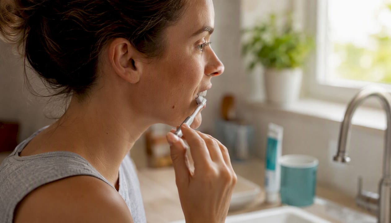
A dentist or periodontist measures recession with a periodontal probe, a thin calibrated instrument that records millimeters of exposed root from the cementoenamel junction (CEJ) to the current gum margin. Alongside that, they record pocket depth, the distance from the gum margin down to where the tissue attaches to the root. Together, these two numbers tell the clinician how much attachment has been lost.
Most clinicians also use recession classification systems to guide treatment planning. The Cairo classification (2011) and the older Miller classification are both referenced in the literature. Cairo’s RT1, RT2, and RT3 categories describe whether the recession is isolated or accompanied by interproximal attachment loss, which directly affects how much root coverage surgery can realistically achieve.
Photographs taken at each visit create a visual record that makes subtle progression visible over time. Radiographs are added when bone loss is suspected, since a two-dimensional X-ray can reveal vertical bone defects that probing alone might miss.
Signs that mean you should not wait
-
Recession that has visibly worsened since your last visit
-
Severe, constant sensitivity that disrupts eating or sleeping
-
A dark spot or soft area on an exposed root (possible root decay)
-
A tooth that feels loose or has shifted position
-
Bleeding, swelling, or pus along the gumline
-
A large exposed root defect covering more than a third of the tooth’s length
General dentist vs. periodontist
Your general dentist can diagnose recession, manage early cases with conservative care, and place composite restorations to cover exposed roots. When surgical grafting is on the table, a referral to a periodontist is standard. Periodontists complete three additional years of specialty training focused specifically on gum and bone conditions, and they perform root coverage procedures routinely. For complex cases involving multiple teeth, bone loss, or combined defects, the periodontist and your general dentist typically coordinate care together.
What non-surgical treatments can stop recession from getting worse?
Conservative care can stop progression and reduce symptoms for many patients, but it does not restore lost gum volume. Think of it as damage control and stabilization, not reversal.
Daily home-care steps
-
Soft-bristled toothbrush only. Medium and hard bristles have no place in a recession-prone mouth.
-
Modified Bass technique. Angle bristles at 45 degrees to the gumline, use short circular strokes, and apply only light pressure.
-
Floss or interdental brushes daily. Plaque between teeth is a primary driver of interproximal recession and periodontal disease.
-
Desensitizing toothpaste. Potassium nitrate (5%) calms nerve activity; stannous fluoride formulations (such as Sensodyne Pronamel or Colgate Sensitive) also occlude dentinal tubules and provide fluoride protection. Use consistently for at least four weeks before judging effectiveness.
-
Alcohol-free antimicrobial rinse (chlorhexidine or cetylpyridinium chloride) when your dentist advises it, particularly after SRP.
-
Smoking cessation. No home-care routine fully compensates for tobacco’s effect on gum tissue perfusion and healing.
In-office non-surgical options
Scaling and root planing (SRP) is the cornerstone of non-surgical periodontal therapy. The hygienist or dentist removes calculus and bacterial biofilm from below the gumline, smooths the root surface, and allows the tissue to reattach or at least stabilize. A common staged approach involves SRP followed by a re-evaluation at roughly two to three weeks, then a decision on whether surgical intervention is needed.
Fluoride varnish applied professionally protects exposed root surfaces from decay and can reduce sensitivity within a single visit.
Composite bonding covers exposed root surfaces with tooth-colored resin. It does not add gum tissue, but it seals the root, reduces sensitivity, and improves appearance. It is a reasonable option when recession is stable and surgery is not indicated or desired.
Nightguard fabrication is non-negotiable when bruxism is a contributing factor. A custom occlusal guard worn during sleep reduces the lateral forces that stress gum attachments.
Desensitizing agents comparison:
| Agent | Active Ingredient | Mechanism | Typical Onset |
|---|---|---|---|
| Desensitizing toothpaste | Potassium nitrate 5% | Nerve depolarization | 4–6 weeks consistent use |
| Desensitizing toothpaste | Stannous fluoride | Tubule occlusion + fluoride | 2–4 weeks |
| In-office fluoride varnish | Sodium fluoride 5% | Tubule occlusion + remineralization | 1–2 visits |
| In-office desensitizer | Oxalate compounds / bonding agents | Tubule occlusion | Same visit |
Pro Tip: Give conservative care a genuine trial of 6–12 weeks after SRP and brushing technique correction before concluding it has failed. Sensitivity often improves significantly in that window. If recession is still progressing at your re-evaluation, that is the signal to discuss surgical options, not before.
What surgical treatments are available for gum recession?
When conservative care has stabilized the disease but significant root exposure remains, or when a patient wants root coverage for aesthetic or protective reasons, surgery becomes the conversation. CAF and tunneling procedures combined with CTG are among the most predictable methods for root coverage; biomaterials offer alternatives with specific tradeoffs.
The main procedures explained
Coronally advanced flap (CAF) repositions existing gum tissue coronally (toward the crown) to cover the exposed root. No donor tissue is harvested. It works best on single or adjacent recessions with adequate tissue thickness and height. Used alone, its predictability is lower than when combined with a graft.
Subepithelial connective tissue graft (CTG) harvests a small piece of connective tissue from the roof of the mouth (palate) and places it under a flap at the recession site. CAF+CTG is the combination most consistently cited in systematic reviews for highest root coverage and long-term stability. The tradeoff is a second surgical site and some palatal discomfort during healing.
Modified coronally advanced tunnel (MCAT) / tunneling procedures create a tunnel under the gum without making vertical releasing incisions. CTG or a biomaterial is threaded through the tunnel and the tissue is advanced coronally. Tunneling preserves blood supply better than traditional flaps and tends to produce excellent aesthetic results, particularly for multiple adjacent recessions.
Free gingival graft (FGG) takes a thicker piece of tissue directly from the palate and grafts it at the recession site. The result is a visible color and texture difference compared to native gum tissue, so FGG is used primarily when the goal is to increase the width of attached, keratinized tissue rather than aesthetic root coverage. It is the go-to when a patient has very little keratinized tissue remaining.
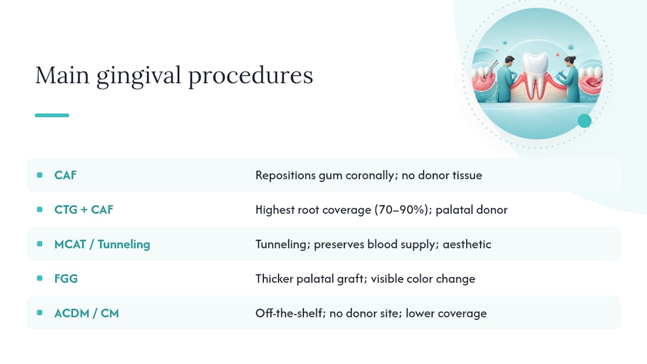
Acellular dermal matrix (ACDM) and collagen matrices (CM) are off-the-shelf biomaterials that substitute for palatal donor tissue. They eliminate the second surgical site and reduce post-operative discomfort. Some multicenter studies report slightly lower root coverage or long-term stability compared to CTG, so clinicians typically counsel patients about that tradeoff. When a patient declines donor-site surgery or palatal tissue is insufficient, ACDM or CM is a well-supported alternative.
Enamel matrix derivative (EMD) (Emdogain) is a protein extract applied to the root surface before grafting to stimulate periodontal regeneration.
Root coverage ranges: Clinical trials and systematic reviews report coverage rates of roughly 70–90% for CTG+CAF procedures, with variability based on tissue phenotype, recession depth, and patient habits such as smoking. Complete root coverage is achievable in many Cairo RT1 cases.
Surgical options at a glance
| Procedure | Best indication | Donor site | Predictability | Aesthetic result | Recovery | Key limitation |
|---|---|---|---|---|---|---|
| CAF alone | Shallow single recession, thick tissue | None | Moderate | Good | 1–2 weeks | Lower alone vs. with CTG |
| CAF + CTG | Single or multiple recessions, moderate depth | Palate | High (70–90%) | Excellent | 2–4 weeks | Palatal morbidity |
| MCAT + CTG | Multiple adjacent recessions | Palate | High | Excellent | 2–4 weeks | Technique-sensitive |
| FGG | Thin tissue, low keratinized band | Palate | High for tissue gain | Fair (color mismatch) | 2–3 weeks | Aesthetic limitation |
| ACDM / CM | Donor-site refusal, limited palatal tissue | None | Moderate | Good | 1–2 weeks | Slightly lower stability vs. CTG |
| CAF + EMD | Adjunct to enhance regeneration | None (adjunct) | Moderate-high | Good | 2–3 weeks | Adjunct only, not standalone |
What should you expect after gum surgery: recovery, risks, and costs?
Recovery timeline
Days 1–3: Swelling, mild to moderate soreness, and some oozing are normal. Cold compresses applied to the cheek (20 minutes on, 20 off) reduce swelling. Stick to soft foods: yogurt, scrambled eggs, mashed potatoes, smoothies. Avoid anything crunchy, chewy, or hot.
Days 4–7: Swelling peaks around day two or three and then gradually subsides. Sutures are typically still in place. Brush only the areas away from the surgical site; use a prescription rinse if provided.
Week 2: Most patients return for suture removal. Sensitivity at the graft site and the palate (if CTG or FGG was used) is still present but improving. Soft diet continues.
Weeks 3–6: The graft integrates and the tissue begins to mature. Color and contour continue to improve. Normal brushing resumes gradually as directed.
8+ weeks: Final aesthetic result is visible. The tissue is stable enough to evaluate coverage percentage. Long-term maintenance appointments are scheduled every three to four months for the first year.
Prognosis and what affects it
Smoking is the single biggest negative factor for graft outcomes. Nicotine reduces blood supply to the graft and significantly increases failure risk. Thin tissue phenotype, deep recession, and poor post-operative hygiene also reduce predictability. Patients with thick tissue, good oral hygiene, and no tobacco use consistently achieve the best coverage results.
Complications to know about
-
Donor site pain (palate): common with CTG and FGG, usually resolves within two weeks
-
Graft failure or partial loss: more common in smokers and patients with thin tissue
-
Infection: rare but possible; signs include increasing pain after day three, pus, or fever
-
Aesthetic mismatch: color or texture difference between graft and native tissue, most common with FGG
-
Recurrence: if the original cause (aggressive brushing, bruxism, periodontal disease) is not controlled, recession can return
Typical U.S. cost ranges
Costs vary considerably by location, provider experience, and whether insurance covers any portion. As a general guide:
-
Periodontal scaling and root planing (SRP): roughly $150–$350 per quadrant, often partially covered by dental insurance
-
Single-tooth connective tissue graft (CTG): approximately $600–$1,200 per tooth out of pocket; some insurance plans cover a portion under periodontal benefits
-
Biomaterial (ACDM/CM) in place of donor tissue: may add $200–$500 to the procedure cost but eliminates palatal surgery
-
Combined restorative + surgical (composite + graft): costs vary based on the extent of the NCCL and the graft procedure chosen
Pro Tip: Ask your periodontist for a pre-authorization from your insurance before scheduling surgery. Many plans cover root coverage grafts when recession is documented as progressing or causing functional problems, even if they exclude purely cosmetic procedures.
When recession coexists with other problems: NCCLs, orthodontics, and bite issues
Recession rarely shows up alone. About 50% of gingival recessions are associated with non-carious cervical lesions (NCCLs), those notch-shaped defects at the gumline caused by abrasion, erosion, or stress fractures. When both are present, the treatment sequence matters.
The NCCL decision tree
If the NCCL is shallow and the root surface is smooth, surgery alone may be sufficient. If the notch is deep, the root surface is irregular, or the defect extends below the anticipated graft margin, the standard approach is to restore the NCCL with composite first, then graft. Placing a graft over an unrestored notch leaves a poor surface for tissue attachment and compromises the aesthetic result. The updated evidence-based recommendation on combined defects provides a structured decision tree for exactly this sequencing question.
Orthodontic considerations
Orthodontic movement can prevent or help correct recession when it repositions roots within the alveolar bone envelope, but it rarely restores tissue that has already been lost. Moving a root buccally through thin bone can create or worsen recession, which is why orthodontists and periodontists often coordinate on patients with a thin biotype before starting treatment. If recession is present before orthodontics begins, a graft may be recommended first to thicken the tissue and reduce the risk of further loss during tooth movement.
Occlusal trauma and bruxism
Lateral forces from grinding or a bite that is off can perpetuate recession even after grafting. An occlusal adjustment (reshaping high spots on the bite) or a custom nightguard is often part of the treatment plan for these patients. Treating only the exposed root without addressing the force causing it is a setup for recurrence.
Interdisciplinary cases, those involving a periodontist, restorative dentist, and sometimes an orthodontist, take longer to plan and execute, but the outcomes are more durable when all contributing factors are addressed together.
How clinicians decide whether surgery is the right call
Understanding the decision process helps you ask better questions at your appointment. Here is the stepwise framework most periodontists use:
-
Identify the etiology. Is the recession caused by periodontal disease, mechanical trauma, thin biotype, orthodontics, or a combination? Treatment that ignores the cause will fail.
-
Control active disease first. No surgery proceeds until periodontal infection is under control. SRP, antimicrobials, and behavior change come before any grafting discussion.
-
Assess tissue phenotype. Thin tissue with minimal keratinized width has a different prognosis than thick tissue. Phenotype influences both technique selection and expected coverage.
-
Evaluate root position and NCCLs. Is the root within the alveolar envelope? Is there a cervical lesion that needs restoration first?
-
Trial of conservative care. For mild recession without active disease, a 6–12 week trial of technique correction and desensitizing therapy is appropriate before committing to surgery.
-
Consider surgical options. If recession is progressing, coverage is desired, or keratinized tissue is critically low, the periodontist selects the technique based on the factors above.
Questions to bring to your consultation
-
How many millimeters of recession do I have, and how does that compare to my last visit?
-
What is my tissue phenotype, and how does it affect my options?
-
What percentage of root coverage can I realistically expect?
-
Do you recommend CTG from my palate, or is a biomaterial appropriate for my case?
-
What does recovery look like, and how long before I see the final result?
-
What does my insurance cover, and what will I pay out of pocket?
-
What happens if I choose not to have surgery right now?
The American Academy of Periodontology and peer-reviewed clinical reviews, including the PMC systematic review cited throughout this article, form the evidence base for these steps. Bringing this checklist to your appointment puts you in a much stronger position to understand the recommendation you receive.
How to prevent further recession and protect your gum health long term
Prevention is not complicated, but it requires consistency. The habits that cause recession develop over years, and so do the habits that stop it.
Daily routine
-
Soft-bristled brush, always. Replace it every three months or when bristles splay, whichever comes first.
-
Gentle, correct technique. Short strokes, light pressure, 45-degree angle to the gumline. Two minutes, twice a day.
-
Floss or use interdental brushes daily. Plaque between teeth is where periodontal disease starts.
-
Desensitizing toothpaste with potassium nitrate or stannous fluoride as your everyday paste, not just when sensitivity flares.
-
Fluoride application. Whether from toothpaste, a prescription gel, or professional varnish, fluoride protects exposed root surfaces from decay.
-
Antimicrobial rinse when recommended by your dentist, particularly after periodontal treatment.
-
Stay hydrated. Dry mouth reduces saliva’s buffering and protective effects on root surfaces.
Behavioral changes that matter
Popular home remedies like oil pulling, essential oils, and lemon juice lack evidence for regrowing gums. At best they reduce surface inflammation temporarily. Skip them and focus on what actually works: eliminating tobacco, reducing acidic food and drink exposure, managing stress (which drives bruxism), and keeping your hygiene appointments at the intervals your dentist recommends, typically every three to four months if you have a history of periodontal disease.
Abrasive whitening toothpastes used daily can accelerate root surface wear. If whitening is a priority, ask about professional options that do not involve daily abrasive exposure.
Pro Tip: At your next hygiene appointment, ask your hygienist to watch you brush for 30 seconds and give you feedback. Most patients have technique habits they are completely unaware of. A single hands-on correction is worth more than any amount of reading about proper brushing. If you use an electric toothbrush with a pressure sensor, the device itself will alert you when you press too hard.
How to choose the right provider for gum recession care
Not every dentist performs root coverage surgery, and not every periodontist has the same depth of experience with all techniques. Asking the right questions before you commit to a treatment plan protects you.
Questions to ask a potential provider
-
How many root coverage procedures do you perform per month?
-
Which techniques do you use most often, and why?
-
Do you offer tunneling procedures (MCAT) as well as traditional flap techniques?
-
What biomaterial options do you have available (ACDM, collagen matrices, EMD)?
-
Can I see before-and-after photos of similar cases?
-
What is your complication rate, and how do you handle graft failures?
-
Do you coordinate with restorative dentists and orthodontists for complex cases?
-
What anesthesia and sedation options are available?
-
How do you handle post-operative pain management?
-
What are your payment and insurance filing options?
Provider checklist
-
Board-eligible or board-certified periodontist for surgical cases
-
Experience with both CTG-based and biomaterial-based techniques
-
Willingness to coordinate with your general dentist on restorative sequencing
-
Clear explanation of expected coverage percentage before you agree to surgery
-
Availability of PRF therapy as an adjunct to support healing, where applicable
-
Transparent discussion of costs and insurance coverage upfront
When reviewing before-and-after photos, look for cases similar to yours in recession depth and location, not just the most dramatic transformations. Ask how long after surgery the “after” photo was taken; tissue continues to mature for months, and a photo taken at six weeks looks different from one at six months.
Our approach to gum recession at Southlittletonfamilydental
At Southlittletonfamilydental, every recession case starts with a thorough evaluation: probing measurements, recession classification, photographs, and a conversation about what is driving the problem. Conservative care comes first. If brushing technique, SRP, or a nightguard can stabilize the situation, that is where we start. When the clinical picture calls for something more, we coordinate surgical planning and restorative sequencing so nothing falls through the cracks. Our general dentistry services include the full range of preventive and non-surgical periodontal care, and our restorative dentistry team handles composite coverage and NCCL restoration as part of a coordinated plan. We believe patients make better decisions when they understand exactly what is happening in their mouth, so we take the time to explain every measurement and every option before anything is scheduled.
Gum recession care is available right here in Littleton, CO
Patients in Littleton and the surrounding area do not need to travel far for a thorough recession evaluation. At Southlittletonfamilydental, a cleaning, exam, and X-ray gives you a complete baseline: recession measurements, pocket depths, photographs, and a clear picture of whether conservative care is enough or whether a surgical conversation is warranted.
New patients can take advantage of the practice’s $99 cleaning package, and an emergency exam is available for $49 when sensitivity or rapid progression cannot wait. The team works with most major dental insurance plans and offers flexible payment options for out-of-pocket procedures. If you have noticed a tooth looking longer, sensitivity that has not gone away, or a visible root surface, the right next step is a scheduled visit with the Southlittletonfamilydental team in Littleton, CO.
What the evidence actually says about treating gum recession
Most articles about receding gums spend a lot of time on home remedies. Oil pulling, aloe vera, green tea rinses. None of them regrow gum tissue. That is not a matter of debate in the clinical literature; it is simply what the evidence shows. The frustrating part is that these remedies are not entirely useless either. Some reduce surface inflammation, which can make gums look slightly healthier for a few weeks. That temporary improvement is exactly what leads people to believe the remedy is working, right up until the next dental appointment reveals the recession has continued.
The more interesting clinical question is not whether surgery works, but when it is actually necessary. The answer is less obvious than most patients expect. A significant number of recession cases, particularly those driven by aggressive brushing in otherwise healthy mouths, stabilize completely with technique correction and professional cleaning. No surgery, no graft, no biomaterial. Just a soft brush used correctly and a hygienist who takes the time to show you how.
Where surgery becomes genuinely necessary is when recession is progressing despite conservative care, when root exposure is causing decay or structural damage, or when the patient has so little keratinized tissue remaining that the gum margin is unstable. In those cases, the evidence for CTG-based procedures is strong and consistent. Systematic reviews conclude that CTG-based procedures generally achieve substantial root coverage across multiple studies and clinical settings.
The piece most patients miss is the maintenance requirement after surgery. A graft that is not supported by correct brushing technique, regular professional care, and bruxism management will eventually fail. The surgery fixes the defect; the habits determine whether it stays fixed.
Gum recession care starts with the right information
Receding gums are not a cosmetic inconvenience. They expose root surfaces that decay faster than enamel, create sensitivity that affects daily life, and, when left unaddressed, can progress to tooth loss. The good news is that the treatment options available today, from simple technique correction to highly predictable surgical grafting, cover the full spectrum of severity.
| Point | Details |
|---|---|
| No spontaneous regrowth | Lost gum tissue does not grow back; treatment stops progression and, when needed, surgically restores coverage. |
| Conservative care first | Soft brush, correct technique, SRP, and desensitizing toothpaste can stabilize mild recession without surgery. |
| Surgery when indicated | CAF+CTG and tunneling procedures achieve 70–90% root coverage in well-selected cases per clinical reviews. |
| Combined defects need sequencing | About 50% of recessions coexist with NCCLs; restoring the notch before grafting improves outcomes. |
| Southlittletonfamilydental in Littleton, CO | Offers recession evaluation, SRP, restorative coordination, and new-patient exam packages starting at $49. |
Bring the clinician checklist from this article to your next appointment. The questions about tissue phenotype, expected coverage percentage, and graft material options are exactly what separates a productive consultation from a vague one.
Sources
These resources are worth bookmarking before your next dental appointment:
Use these sources to frame specific questions for your clinician rather than to self-diagnose. Recession classification, tissue phenotype, and surgical indication all require a clinical examination; no article or website substitutes for that.
This article is general information, not a substitute for advice from a qualified doctor. Consult a qualified healthcare professional about your own circumstances before acting on anything here.