
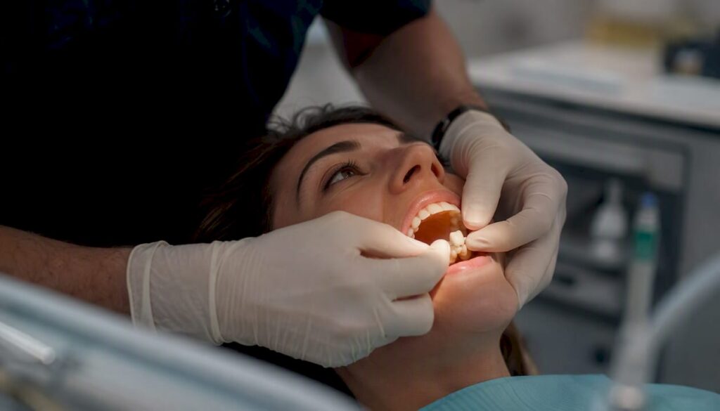
Yes, a tooth can absolutely decay under a dental crown. The crown protects the visible surface, but the underlying tooth structure and the margin where crown meets gum remain vulnerable to bacteria. Decay under a crown, clinically called secondary caries or caries associated with restorations (CARS), is one of the most common reasons crowns fail.
If you suspect a problem right now, here is what to do:
- Call your dentist today if you have pain, swelling, fever, a foul taste, or your crown has come off.
- Rinse gently with warm salt water to reduce irritation, but do not attempt to re-cement a fallen crown at home with store-bought adhesive.
- Avoid chewing on that side and skip very hot, cold, or sweet foods until you are seen.
At your appointment, your dentist will likely perform a clinical exam, take bitewing or periapical X-rays, and may order a cone-beam CT (CBCT) scan if standard imaging is inconclusive. Depending on what they find, treatment could range from a simple marginal repair to crown removal, root canal therapy, or, in severe cases, extraction.
Key Takeaways
Tooth decay under a crown is common, often silent in early stages, and almost always more manageable when caught at a routine exam rather than after symptoms appear.
| Point | Details |
|---|---|
| Decay under crowns is real | The margin where crown meets tooth is vulnerable to bacteria regardless of crown material or age. |
| Early detection changes outcomes | Routine bitewing X-rays catch most cases before root canal therapy or extraction becomes necessary. |
| Imaging has limits | Artifact-free CBCT accuracy is — vs. — for standard X-rays, but drops to — near amalgam. |
| Treatment follows decay depth | Small marginal decay may need only a repair; deep decay reaching the pulp requires root canal plus a new crown. |
| Southlittletonfamilydental | Offers same-day emergency exams, crown evaluation, restorative care, and root canal therapy in Littleton, CO. |
Table of Contents
- How does decay get under a dental crown?
- What are the signs of decay under a crown?
- How do dentists detect decay under crowns?
- What are the treatment options for decay under a crown?
- What happens if you leave decay under a crown untreated?
- How can you prevent decay under a crowned tooth?
- When should you see a dentist about a crowned tooth?
- How Southlittletonfamilydental evaluates and treats crowned teeth
- Southlittletonfamilydental: same-day care for crown concerns in Littleton, CO
- A note on realistic expectations and individualized care
- Sources
How does decay get under a dental crown?
A crown does not fuse to your tooth like a permanent seal. It sits over the prepared tooth structure and is held in place by dental cement, and that junction at the gum line is where trouble typically starts.
The primary mechanical pathway is microleakage: microscopic gaps between the crown margin and the tooth allow bacteria, saliva, and acids to seep in. Over time, those bacteria digest the tooth structure underneath, producing the same acid-driven decay process as any cavity. Marginal gaps above a certain small size are considered clinically significant for secondary caries risk, while modern CAD/CAM restorations tend to achieve tighter fits within an acceptable smaller range.
Several factors accelerate this process:
- Cement breakdown. Older zinc phosphate or polycarboxylate cements can dissolve gradually, widening the marginal gap and opening a direct bacterial pathway.
- Poor marginal fit. A crown that was not perfectly adapted at placement starts with a larger gap, giving bacteria an earlier foothold.
- Crown age. Even a well-fitted crown develops microgaps over years of chewing forces and thermal cycling from hot and cold foods. The seal is dynamic, not permanent.
- Pre-existing decay. If decay was not fully removed before the crown was placed, bacteria remain trapped underneath and continue progressing.
- Gum recession. When the gum pulls back, it exposes root surface below the crown margin, an area with thinner enamel and higher cavity risk.
- Dry mouth. Saliva neutralizes acids and washes away bacteria. Reduced flow from medications or systemic conditions removes that protection.
- Bruxism. Grinding stresses the crown margins repeatedly, accelerating cement fatigue and microcracking.
- High-sugar or high-acid diet. Frequent acid exposure keeps the oral environment hostile to tooth structure, especially at vulnerable margins.
Pro Tip: If your dentist has noted bruxism, a custom night guard does more than protect your teeth from wear. It reduces the cyclical stress on crown margins that speeds up cement breakdown. Pair it with a prescription-strength fluoride toothpaste or a fluoride varnish applied at checkups, and you meaningfully slow the conditions that lead to marginal decay.
What are the signs of decay under a crown?
The frustrating reality is that early decay under a crown often produces no symptoms at all. Many cases are caught only on routine X-rays, which is exactly why those checkups matter.
Early signs (often subtle):
- New sensitivity to hot, cold, or sweet foods that was not there before
- A bad taste or odor near the crowned tooth, sometimes described as a crown smells bad situation caused by bacterial activity at the margin
- Bleeding or swollen gum tissue right at the crown margin
- Floss shredding or catching at the edge of the crown
Later signs (indicating more advanced disease):
- Persistent, throbbing pain that lingers after the temperature stimulus is gone
- Crown mobility or a feeling that the crown has shifted
- Visible dark staining or discoloration at the gum line where crown meets tooth
- Pus, visible swelling, or a pimple-like bump on the gum near the tooth
Decay under a crown can be completely silent until it reaches the pulp. By the time pain becomes constant or swelling appears, the infection has typically progressed well beyond the margin. Routine X-rays catch the majority of cases while they are still manageable, before root canal therapy becomes necessary.
Seek same-day care immediately if you experience: severe spontaneous pain, facial swelling, fever, difficulty swallowing, or if the crown falls off and the exposed tooth is painful. These are not “wait and see” situations.
How do dentists detect decay under crowns?
Diagnosing decay beneath a crown is genuinely difficult. Crown materials, especially metal, block or distort X-ray beams, which can hide decay or create false shadows. Here is the typical stepwise approach:
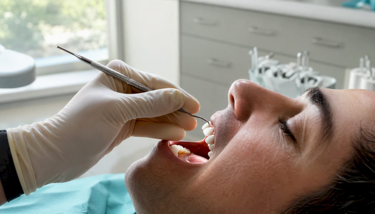
Clinical inspection and probing. The dentist examines the crown margin visually and uses a fine probe to check for softness, gaps, or bleeding. This catches obvious marginal breakdown but cannot reveal what is happening deeper under the crown.
Bitewing and periapical X-rays. These are the standard first-line imaging tools. They can reveal decay at or near the margin, bone changes, and pulp involvement. The limitation is that dense crown materials, particularly metal-ceramic and full-metal crowns, produce radiographic artifacts that obscure the underlying tooth structure. A cavity can hide directly behind the crown shadow.
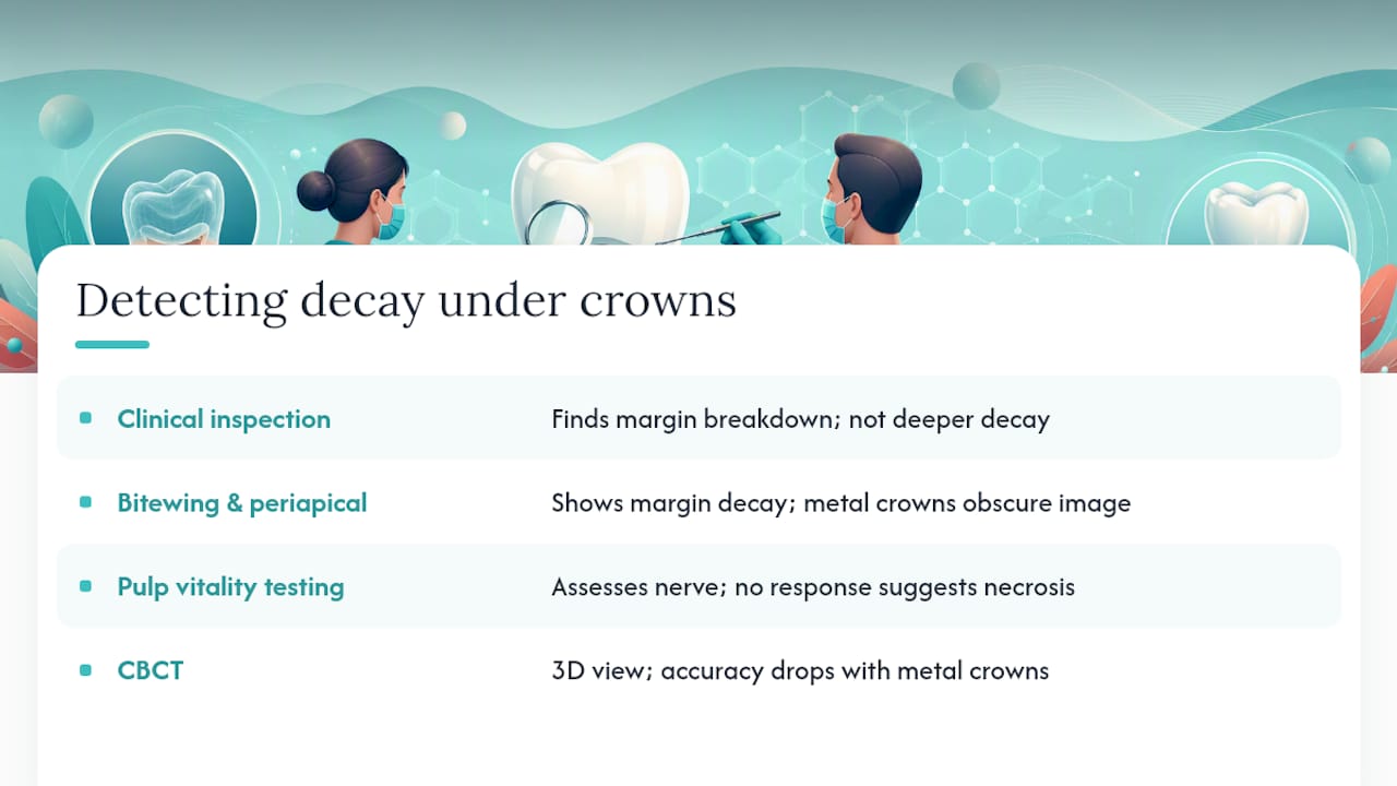
Pulp vitality testing. Cold tests or electric pulp testers assess whether the nerve is still responding normally. A tooth that no longer responds may have pulp necrosis, which changes the treatment path significantly.
CBCT imaging. Cone-beam CT provides a three-dimensional view and can improve detection when standard X-rays are inconclusive. However, accuracy varies considerably depending on what the crown is made of.
Research published in a Springer study on diagnostic accuracy comparing CBCT and intraoral radiography found the following:
The takeaway: CBCT is more accurate than standard X-rays when there is no interfering material nearby, but its accuracy actually drops below that of conventional radiography when the crown contains amalgam or dense metal. An “artifact” in this context means a streak or shadow created by the dense material that the scanner misreads as tooth structure, or that masks real decay.
When imaging remains inconclusive, dentists sometimes remove the crown entirely for direct visual inspection of the underlying tooth. That is not a failure of technology; it is the clinically appropriate response when the stakes are high and the picture is unclear.
Pro Tip: Ask your dentist which crown material you have. If it is a full-metal or metal-ceramic crown, mention it before imaging is ordered. That detail affects which imaging protocol gives the most useful information and whether CBCT is worth the added cost and radiation dose in your specific case.
What are the treatment options for decay under a crown?
Treatment follows a logical progression based on how far the decay has spread. The earlier it is caught, the more conservative the fix.
Decision flow:
-
Small marginal decay (tooth structure mostly intact). The dentist may repair the cavity at the margin without removing the crown, using composite resin or glass ionomer cement to seal the area. This works when the decay is accessible and the existing crown is otherwise sound.
-
Moderate decay (crown must come off). The crown is removed, decay is excavated, and the tooth is rebuilt with a core buildup. If the existing crown is undamaged and still fits well, it may be recemented. More often, a new crown is fabricated because the tooth shape has changed after decay removal.
-
Deep decay reaching the pulp. Root canal therapy is needed before any crown work. The canals are cleaned and sealed, a post and core may be placed, and a new crown is made. This is a multi-visit process, typically two to four appointments over several weeks, with a temporary crown in place between visits.
-
Non-restorable tooth. When decay has destroyed too much tooth structure or the root is compromised, extraction is the only option. Replacement choices include a dental implant, a fixed bridge, or a partial denture.
Pros and cons at a glance:
- Marginal repair: Least invasive, lowest cost, preserves existing crown. Only works for small, accessible lesions.
- Crown removal and replacement: Addresses the full extent of decay, allows direct inspection. Requires a new crown and temporary restoration period.
- Root canal plus new crown: Saves the tooth when pulp is involved. Multiple visits, higher cost, but avoids extraction.
- Extraction plus implant: Permanent replacement, no ongoing decay risk in that tooth. Longest timeline and highest upfront cost; the implant process typically spans several months.
Catching decay early almost always means a simpler, less expensive path. A marginal repair costs a fraction of a root canal and new crown, and a root canal costs far less than extraction followed by implant placement.
What happens if you leave decay under a crown untreated?
Decay does not pause on its own. Without treatment, the progression follows a predictable and increasingly serious path:
- Bacteria at the margin erode through the outer dentin layer.
- Decay reaches the inner dentin, causing sensitivity and pain.
- Bacteria enter the pulp chamber, triggering pulpitis (nerve inflammation).
- The pulp dies, and infection spreads into the root canal system.
- An abscess forms at the root tip, with bone loss in the surrounding jaw.
A dental abscess is a medical emergency. Untreated oral infections can spread to the jaw, neck, and in rare cases the airway or bloodstream. If you notice facial swelling, fever, or difficulty breathing alongside dental pain, seek emergency care immediately.
Signs that more invasive treatment is likely already needed:
- Constant, unprovoked pain that wakes you at night
- Swelling of the gum, jaw, or face near the tooth
- A bad taste that persists despite brushing, indicating pus drainage
- The crown feels loose or has shifted noticeably
- Fever accompanying dental pain
Beyond the biological risks, delayed treatment consistently means higher costs. A marginal repair that might have cost a few hundred dollars becomes a root canal, post, and new crown. If the tooth is lost, replacement with an implant adds months of treatment and significantly more expense.
How can you prevent decay under a crowned tooth?
A crown is a restoration, not a guarantee. The tooth underneath still needs the same care as every other tooth, and the margin needs a little extra attention.
Daily care checklist:
- Brush twice daily, angling the bristles at 45 degrees toward the gum line to clean the crown margin directly. An electric toothbrush with a pressure sensor reduces the risk of brushing too hard while improving plaque removal.
- Floss daily around the crown. If standard floss is difficult, try a floss threader, interdental brushes, or a water flosser. The goal is disrupting plaque at and just below the margin.
- Use a fluoride toothpaste. For higher-risk patients (dry mouth, history of frequent cavities, multiple crowns), a prescription fluoride toothpaste or a fluoride rinse adds meaningful protection at the margin.
- Limit frequent snacking on sugary or acidic foods. Every acid exposure is a small attack on the margin; spreading meals out gives saliva time to neutralize and remineralize.
Professional maintenance:
- Routine exams and X-rays every six months remain the most reliable way to catch early decay under crowns before symptoms appear. Bitewing X-rays at these visits are specifically designed to show marginal areas.
- Ask your dentist about fluoride varnish applied at cleanings, particularly if you have multiple crowns or a dry mouth condition.
- If you grind, wear your night guard consistently. Bruxism accelerates cement breakdown at margins, and a well-fitted guard is one of the most cost-effective protective measures available.
Pro Tip: The cement type used when your crown was placed affects long-term marginal integrity. Adhesive resin cements and resin-modified glass ionomer cements generally provide better marginal sealing than older zinc phosphate cements. If you are having a crown replaced, ask your dentist which cement they plan to use and why.
When should you see a dentist about a crowned tooth?
Same-day or emergency visit (do not wait):
- Crown has fallen off and the exposed tooth is sensitive or painful
- Severe, constant pain that is not relieved by over-the-counter pain medication
- Visible swelling of the gum, jaw, or face
- Fever accompanying dental pain
- Pus or a bad taste that returns after rinsing
If your crown falls out: keep it clean and bring it to your appointment. Store it in a small container with a little water or milk. Do not try to glue it back with household adhesive or super glue. The exposed tooth is vulnerable to temperature, pressure, and bacteria, so getting seen the same day matters.
Routine visit (book within a week or two):
- New sensitivity to hot, cold, or sweets that was not present before
- Floss consistently shredding or catching at the crown edge
- Mild, intermittent discomfort that comes and goes
- Visible dark line or staining at the crown margin
- Crown that feels slightly different when you bite but is not painful
At a same-day emergency visit, expect a targeted exam, X-rays, and a treatment decision or temporary stabilization that day. A routine visit allows more time for comprehensive imaging and a staged treatment plan.
How Southlittletonfamilydental evaluates and treats crowned teeth
When a patient comes in with a concern about a crowned tooth, the evaluation at Southlittletonfamilydental follows a structured clinical workflow designed to get a clear picture before any treatment decision is made.
What happens at the exam:
- A thorough history: when symptoms started, what makes them better or worse, any recent changes to the crown
- Targeted visual and tactile examination of the crown margin, checking for gaps, staining, or soft spots
- Bitewing and periapical X-rays to assess the margin, surrounding bone, and pulp
- Pulp vitality testing to determine nerve status
- CBCT imaging when standard X-rays are inconclusive, interpreted in the context of the crown material to account for artifact risk
- A clear treatment estimate with options explained before any procedure begins
Patients deserve to understand what is happening in their own mouth. At Southlittletonfamilydental, the goal at every appointment is to explain what the imaging shows, what the options are, and what happens if treatment is delayed — so patients can make informed decisions without pressure.
Relevant services for crowned-tooth issues at the practice include restorative dentistry (crown replacement, core buildups, fillings), root canal therapy, extractions, and same-day emergency exams. When you call to schedule, bring your insurance information and any previous X-rays if you have them. Recent images from another office can save time and reduce the need for repeat imaging.
Southlittletonfamilydental: same-day care for crown concerns in Littleton, CO
Worried about a crowned tooth? At Southlittletonfamilydental, a same-day emergency exam starts at $49, and the new patient cleaning, exam, and X-ray package gives you a full diagnostic baseline without the sticker shock. The practice uses advanced imaging to evaluate crown margins accurately, and every treatment plan is explained in plain language before anything is scheduled.

Whether you need a crown evaluated, a root canal, or a full crown replacement, the team handles it all in one location. Patients facing tooth replacement after extraction can also discuss implant options at the same visit. Book your appointment at Southlittletonfamilydental and bring any previous X-rays to get the most out of your first visit.
A note on realistic expectations and individualized care
Every crowned tooth has its own history, and no two cases follow exactly the same path. Some patients come in with a crown that has been in place for fifteen years and is still holding strong. Others find decay at the margin within a few years of placement, often because of dry mouth, grinding, or a diet that keeps the oral environment acidic.
What matters most is that monitoring does not stop once a crown is placed. Crowned teeth need the same routine attention as natural teeth, and in some ways they need more, because the margin is a permanent structural vulnerability that requires ongoing observation. Multi-stage treatments like root canal therapy followed by a new crown take time, and patients should expect clear communication at each step about what comes next and why.
This article is general information, not a substitute for advice from a qualified doctor. Consult a qualified healthcare professional about your own circumstances before acting on anything here.
Sources
- Diagnostic accuracy of CBCT and intraoral radiography for detection of proximal caries adjacent to different restorative materials (Springer)
- Imaging challenges and interpretation issues for restored teeth (PMC article)
- Etiology of Secondary Caries in Prosthodontic Treatments | IntechOpen